Showing posts with label disease. Show all posts

Monday, October 24, 2011

October 24th, 2011


Today, we started class by receiving a sheet on some new requirements for our blogs. They are that each blog you do must have at least one picture, and you must comment of other peoples' blogs st least 3 times before each person has finished the 2nd round, but cant do more that 2 comments a week. These comments could include constructive criticism as long as actually productive, and comments like "blah blah blah... was really good" are not allowed and aren't useful, so you should think before posting a comment.
After that, we finished our notes on what makes us sick, but because the pages were mixed up we sort of had to skip around. We did pages (not in the correct order by the way) 22, 24, 1st half of 25 but other half was a review of T and B cells, 26, 27, 28, and 29. these pages were focused on the T and B cells, and how they worked. T cells have 3 types, cytotoxic (killer) T cells, helper T cells, and a third type (found in the study section on pages 49-55 which you should read) called Suppressor T cells. Helper T cells identify the foreign substance in the body, mark it to be destroyed, and stimulates the growth of cytotoxic T cells and B cells. Cytotoxic T cells kills infected body cells that are malfunctioning or are producing pathogens. Suppressor T cells slows activity of T and B cells after the infection is dealt with. B cells produce memory cells and plasma cells. Plasma cells create antibodies to combat the infection and memory cells keeps formula of cells that combat a certain infection or disease.
We also learned about primary and secondary immune responses. The primary immune response occurs when a new or mutated pathogen enters the body, and it takes a few days to produce antibodies, but the memory cells store formula to combat re-infection. Secondary immune response occurs with the pathogens 2nd infection, and it killed off much more rapidly because of the memory cells, and is often symptom free.

Immune disorders were in our notes as well. they are the consequence of a malfunction of the immune system. They include allergies, autoimmune disorders - system turns against bodies own molecules, and immunodeficiency diseases - when body lacks one of more parts of the immune system. Some types of autoimmune diseases are rheumatoid arthritis, juvenile diabetes, multiple sclerosis, and lupus. Some immunodeficiency diseases are SCID (Severe Combined Immunodeficiency) which there is few T and B cells, Hodgkin disease, and AIDS/HIV, which attacks helper T cells.
After that we watched a few short movies on T and B cells/antibodies. Antibodies attach to pathogens, stopping them from infecting cells (neutralization), then preform agglutination, or clumping so a phagocyte can kill them in phagocytosis. B cells make humoral immunity, in which B cells send out antibodies, each which can only bind to one type of antigen and make memory cells and plasma cells. Helper T cells sends signals to stimulate growth of other T and B cells after marking infected cell.

HW: Spice lab, Read UP pages49-55, do UP pages 45-46
Next scribe: Kiran

Sunday, October 23, 2011

October 21st, 2011


Assignments due for today:
Bacteria labs analysis
pp.31-32 in the UP packet (Identifying Agents of Disease)--review for friday's test!!!
Assigned today:
Spice lab I-V (data tables)--10/25
Read Chp. 24 p.528-543 (complete backside of wksht)
Disease video due 10/31
Study for test on 10/28!!
Received a "study guide" for bacteria and viruses

Today in class, we watched a germ theory video and filled in our note packet up to page 23.

The germ theory is that a microorganism causes a disease (germs).

Be familiar with:
diptheria tuberculosis mumps
the common cold whooping cough pneumonia
influenza poliomyletis meningitis
malaria rabies ringworm
typhus infectious mononucleosis tetanus
chicken pox AIDS Rocky Mountain spotted fever
amebic dysentery streptococcal sore throat German measles
Botulism Athletes foot measles (rubeola)

Some key points in our packet:

  • Nonspecific defenses (when the body doesn't distinguish one infectious microbe from another):

First line of defense= 1. skin
2. Mucous membranes (ex. eyes water)
3. secretions of skin and mucous membranes
Second line of defense= 1. Phagocytic (cell-eating) white blood cells
(part of the lymphatic 2. Defensive proteins
system) 3. The inflammatory response
  • Specific defenses (immune system--when the body recognizes the pathogen):
Third line of defense= 1. Lymphocytes (white blood cells)
(part if the lymphatic 2. Antibodies
system)

A. Nonspecifics have two types of defense: External and internal.
Internal defenses are like the external's backup--they defend when an intruder gets by
the external barriers.
A type of internal defense is an interferons: they slow/stop viral replication
Where an infected cell "calls for help" and a neighboring cell sends proteins to inhibit
viral replication.

The inflammatory response= when tissue is damaged.
You know when the immune system is working when the injury (such as a small cut)
becomes red, swollen (because of blood vessels dilating) , and warm to the touch.
The lymphatic system consists of a network of vessels (similar to those of the circulatory system) and lymph nodes.
Its two main functions are to return tissue fluid to the circulatory system and to fight infection. It is the main "battle ground" for when the body is fighting infection.

B. Specific defenses: for when nonspecifics fail.

Immune system recognizes and attacks specific intruding microbes:
bacteria pollen parasitic worms
cancer cells house dust cells of transplanted tissue
protozoa mold spores
(all allergies)

Key players= antigens (elicit immune response) and antibodies (proteins found in blood plasma--mark invaders: neutralization and agglutination)
Antibody
Variable portion= there are two variable portions to each antibody so they can latch on to two different invading microbes. If enough accumulate and "clump" together (agglutination), the microbes cannot function.

STUDY FOR TEST:

Notes packet
bacteria/virus sheet (p. 31-32 in UP)
"study guide" handed out in class

NEXT SCRIBE: Sean


Thursday, October 20, 2011

OCTOBER 20, 2011


Today in class, we completed page 33 in the UP, pages 12-15 in the notes, and finised the bacteria lab.
Page 33: Analyse the diagram provided in the UP and answer the subsequent questions. Correct answers to the questions posted below.
1. Which of the antibiotics would you use to prevent the growth of B. subtilis?
Neomycin is the best antibiotic for inhibiting B. subtilis growth. Aureomycin and erythromycin also work.
2. Which of the antibiotics would you use to prevent the growth of E. coli?
Tetracycline is the best antibiotic for inhibiting E. coli growth. Aureomycin also works.
3. Are both organisms equally sensitive to antibiotics? Explain.
No. More antibiotics inhibit one organism, with greater effect, than the other.
4. Which of the two organisms are more sensitive to antibiotics in general?
B. Subtilis
5. If you wanted to inhibit both organisms with one antibiotic, which would you use?
Aureomycin
6. If E. coli is beneficial and B. subtilis is harmful and you were infected with both, which antibiotic would you use?
Neomycin would inhibit the growth of both bacteria best. Erythromycin would also work.
7. In general, what can you conclude about bacteria and antibiotics from this experiment?
Antibiotics are not as specific as enzymes, and may inhibit the growth of multiple strains of bacteria. Different bacteria are sensitive to different antibiotics with different degrees of sensitivity.
8. What features does this experiment lack that it should have?
A control group
9. How would you correct this omission?
Add a paper disk.
Notes:
Bacteria Lab:
Surface Lab:
1. Find your petri dish. DO NOT open it! Possibly dangerous bacteria could be growing in it.
2. Observe the petri dish. You should see bacteria colonies (they look like little circular clumps). Count the number of colonies present on the surface of the agar. Do not confuse bacteria colonies with fungus, which also may have grown. Fungus will have little "arms." Don't count it!
3. Record the number of colonie in each quadrant.
4. Safely dispose of the petri dish.
Antibiotic lab:
1. Find your petri dish and a metric ruler.
2. Find the zones of inhibition around each antibiotic, if any. They should look like clear, bacteria free circular "halos' around the antibiotic disk.
3. Measure the diameter of each zone of inhibition in millimeters. If the circle of the zone of inhibition is not complete or fully measurable, measure the radius and multiply it by two.
4. Record the diameter of each zone of inhibition for each antibiotic.
5. Safely dispose of the petri dish.
Results of these labs varied between groups. If you were absent today and unable to recive results from your group, here is a sample of some of the results collected today:
Surface Lab:
Control: 12 colonies
Doorknob: 145 colonies
Faucet Handle: 78 colonies
Desk: 178 colonies
Antibiotic Lab:
Control: 0 mm
Streptomycin: 24 mm
Penicillin: 13 mm
Neomycin: 15 mm
Homework: Continue working on reasearch/ scripts for disease project, finish pp. 31-32 in UP for tomorrow, and begin spice lab (pp. 37-41 in UP)
NEXT....Emma

Wednesday, October 19, 2011

October 19, 2011

Today, we set up our Bacteria Labs in class so that they would ready for us to observe the following day. We split up into groups of two. One of the partners set up "How common are bacteria and how quickly do they reproduce"
They needed to follow the directions on pg 19-20 in UP to do this which included

labeling their dish into 4 separate areas
then using 3 different items to wipe on top of the different areas, one in each, with no object wiped in the fourth section as that is the control
make sure to label which items were wiped in each section
close your petri dish now and give it to your teacher

The other partner followed the procedure on pages 23-24 for Lab "Using Antibiotics to stop bacterial growth"
follow these steps:
label your dish into 4 separate areas
take a cotton swab, dip it in bacteria broth, and then wipe it all of the the petri dish nutrient agar so that each section has the same amount
take three different antibiotics, using the tweezers, and place them in separate sections
take a plain piece of paper and put it in the fourth section as your control
make sure you label which antibiotic is in each section
close your petri dish now and give it to your teacher


We also took an extensive amount of notes, all of which can be found through the Gbs Moodle page if you go to the Bio Metacourse and click on notes for UNIT 3. For an odd reason, my link is not working however so I hope that I am just unlucky and that you will all have no trouble with the link loading. The pages we covered in the notes packet were 1-12.

HW:
Read "Just an Upset Stomache" pg 27-28 and highlight key points
Read pg 29 as well


Tuesday, October 18, 2011

OCTOBER 18TH 2011

Today in class we watched a video and filled out a sheet that was aimed at inhancing our understanding of bacteria.
The work sheet went over the biological weapon botualism which was created by bacteria.


Botulism Food Poisoning
http://www.schmidtandclark.com/botulism

TO YOUR LEFT IS THE BOTULISM VIRUS


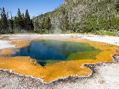
The video also discussed that bacteria can live in the harshest environments including hot spings, ocean vents and caves.

http://www.google.com/imgres?q=bacteria+in+hot+springs&num=10&hl=en&gbv=2&biw=1280&bih=596&tbm=isch&tbnid=51JI8a05XgVdnM:&imgrefurl=http://www.biofpr.com/view/MTA2NDQzL0FSLzUxL251bGw%3D/featureDetail.html&docid=PRqbX1mavxNBzM&imgurl=http://www.biofpr.com/SpringboardWebApp/userfiles/biofpr/image/features/08/features_0809_hot_springs.jpg&w=240&h=180&ei=HfedTqvaBtPciAKE0OHiCQ&zoom=1&iact=hc&vpx=891&vpy=165&dur=2212&hovh=144&hovw=192&tx=99&ty=86&sig=101603448198120267361&sqi=2&page=1&tbnh=116&tbnw=155&start=0&ndsp=19&ved=1t:429,r:4,s:0

This picture above shows bacteria that has entered and found its way in a hot spring at Yellowstone National Park.


The video also went over where bacteria can be found in our bodies and how they are benficial to us humans.


Bacteria is found on our mouths and is on your mouth as you are reading this.


http://www.google.com/imgres?q=bacteria+on+mouth&hl=en&sa=X&gbv=2&tbas=0&biw=1280&bih=596&tbm=isch&tbnid=toS6tz3E1uXmEM:&imgrefurl=http://medicineworld.org/main-archives/Aug-10-2007.html&docid=EVdV7Y1QC8vDIM&imgurl=http://medicineworld.org/images/blogs/thumbs/7-2007/mouth-bacteria-12981-thumb.jpg&w=120&h=158&ei=g_mdTpOtL8rqqQGcrLiKDw&zoom=1&iact=hc&vpx=949&vpy=184&dur=718&hovh=125&hovw=94&tx=78&ty=55&sig=101603448198120267361&page=1&tbnh=119&tbnw=87&start=0&ndsp=21&ved=1t:429,r:5,s:0


The picture above reveals a portion of someones tongue and zoomed in on it and found bacteria. The bacteria on our


tounges help us digest our food .


The video brought up an interesting topic. It stated that sourdough bread gets its flavor from bacteria from the mother


dough.


http://www.google.com/imgresq=sourdough+bread&hl=en&sa=X&gbv=2&tbas=0&biw=1280&bih=596&tbm=isch&tbnid=EjTqUtqFtqmVSM:&imgrefurl=http://www.truestarhealth.com/Notes/1940007.html&docid=PdrJ322fLBm0gM&imgurl=http://www.truestarhealth.com/Notes/Images/Food_Guide/Sourdough_Bread.jpg&w=200&h=200&ei=4_qdTqGWCYLNqgGd74TKDg&zoom=1&iact=rc&dur=509&sig=101603448198120267361&page=1&tbnh=126&tbnw=128&start=0&ndsp=18&ved=1t:429,r:3,s:0&tx=57&ty=55


The video also discussed how streptococcus bacteria can infect many different areas in our


bodies and feeds on the tissue within the area its infecting.


http://www.google.com/imgres?

q=streptococcus&hl=en&gbv=2&biw=1280&bih=596&tbs=isz:m&tbm=isch&tbnid=xReZPEyfvWqs3M:&imgrefurl=http://student.ccbcmd.edu/courses/bio141/lecguide/unit2/bacpath/plaque_SEM.html&docid=CheQR8nTkBnCmM&imgurl=http://faculty.ccbcmd.edu/courses/bio141/lecguide/unit2/bacpath/images/plaque_tooth.jpe&w=414&h=296&ei=j_ydTqueHorcqgGfvuTKDg&zoom=1&iact=hc&vpx=847&vpy=233&dur=1563&hovh=190&hovw=266&tx=162&ty=102&sig=101603448198120267361&page=3&tbnh=157&tbnw=193&start=31&ndsp=10&ved=1t:429,r:3,s:31


This is streptoccoccus growing on the enamel of a tooth.


We also learned that Alexander Flemming accidently discovered a mold that killed bacteria and


later isolated the mold and created penicillin.


http://www.google.com/imgres?

q=penicillin+pills&hl=en&sa=X&gbv=2&tbas=0&biw=1280&bih=596&tbm=isch&tbnid=yZAhq2_F0ApW9M:&imgrefurl=http://www.rxzone.us/product.cfm/rx/penicillin-


vk-250-mg-tablets-each-7


37460.html&docid=EqRG8w914O66zM&imgurl=http://www.rxzone.us/images/products/big/737460.jpg&w=288&h=216&ei=5_6dTv3pBIjTqgHTq_jJDg&zoom=1&iact=hc&vpx=364&vpy=312&dur=1532&hovh=172&hovw=230&tx=122&ty=116&sig=101603448198120267361&page=1&tbnh=123&tbnw=164&start=0&ndsp=13&ved=1t:429,r:8,s:0


This is a penicillin pill ment to be taken orally.


Antibiotics are active against a diseases.


One of the main points that i got out of this movie was how traveling can affect the spreading of


bacterial infections. I learned that especially international travelers are constantly bringing in


new bacteria from the where they came from and are immune to the bacteria that already exist in


the place that they are visiting.


NEXT SCRIBER IS JACK

Monday, October 17, 2011

October 17, 2011

AGENDA-
Today in class we finished what was left of the Flu video from October 13th. The last part of the video basically summarized how Holly Jones was recovering from her influenza virus with antibodies, etc.


"Travel inside the body of Holly Jones, a 25-year-old motorcycle courier who becomes a victim in the cellular war between her immune system and the influenza virus."

http://school.discoveryeducation.com/lessonplans/programs/operationantibody/q.html


After we finished the last part of the previous video, we started a new film "Understanding Viruses" (part 1 and most of part 2), along with the questions on page 15 of the Unit Packet. During the film, we were to answer the questions, turning in p.15 of UP at the end of class.


The video briefly talked about an important virologist, Jonas Salk. He was famous for creating an effective Polio Vaccine.


http://www.famous-scientists.net/jonas-salk.htm


"Understanding Viruses" Video- Part 1/17:

Part 2/17: http://www.youtube.com/watch?v=kq9thrI6zlo&feature=related
Part 3/17: http://www.youtube.com/watch?v=sUHrGD89Db4

*For parts 4-17, search "Understanding Viruses part (#)/17" into youtube.com

While students watched the "Understanding Viruses" video, Mrs. Andrews showed us our grades for the Ecology in Your Backyard project.

HOMEWORK-
•Read CH 15 p. 303-309 (with note sheet)
•Read UP p. 19-25 - bacteria labs
•Fill in UP p. 31-32 (use text / internet) - Due Friday, 10/21

NEXT SCRIBE-
Jimmy

Thursday, October 13, 2011

October 13, 2011

Today in class, we went through the notes about viruses and watched a video about the flu. Here's a basic overview of the notes.
Viruses: Living or non-living?
  • viruses are between life and non-life
  • Living characteristics: contain genes (RNA or DNA), are highly organized (complex protein structure
  • Non-living characteristics: not made of cells, cannot reproduce on thier own: viruses don't have the necessary structures to reproduce so they must use the structures of living cells

Structure

  • nucleic acid, the genetic material, is inside a protein coat
  • often include: head (where genes are held), tail, tail fibers

The virus in the animation on the left is called a T4 Bacteriophage. It is easy to see the structure of this virus.

http://www.emc.maricopa.edu/faculty/farabee/biobk/biobookdiversity_1.html

Reproduction: Lytic and Lysogenic Cycles

Lytic Cycle

  1. virus injects genes into a host cell
  2. "hijacks" cell structures and uses them to make copies of itself
  3. viruses eventually causes the cell to lyse (burst)
  4. viruses are released to find a new host cell

Lysogenic Cycle

  1. virus injects genes into host cell
  2. viral genes are incorporated into normal cell genes
  3. genes are passed on without the cell making copies of the virus
  4. while in lysogenic cycle, cell does not lyse
  5. environmental changes will trigger the viral genes to switch to the lytic cycle

http://terra.dadeschools.net/Books/Biology/BiologyExploringLife04/0-13-115075-8/text/chapter16/concept16.5.html

Plant Viruses

  • affect plants: interfere with growth and damages crops
  • most plant viruses have RNA not DNA
  • viruses are designed to get past plant epidermis and cell walls
  • no cure for the majority of plant viruses
  • humans have created plants that are resistant to certain viruses

Examples: Tobacco mosiac virus (TMV) and tomatoes, PRSV and papaya

Animal Viruses

  • many have a phosolipid outer layer, just like a cell's plasma membrane: allows viruses to slip in and out of the cell through endocytosis and exocytosis
  • reproduce in cell's cytoplasm: needs ribosomes to make viral proteins
  • RNA and DNA viruses
  • RNA viruses: HIV, mumps, common cold, polio
  • DNA viruses: chicken pox, herpes, hepatitis

http://www.sciencegateway.org/resources/biologytext/cb/virus/virus.html

HIV: the cause of AIDs

  • Retrovirus: reproduces by the use of viral DNA molecules
  • normally, cells use DNA to produce RNA (transcription)
  • HIV has cells use RNA to produce DNA (reverse transcription)
  • RNA then makes proteins
  • viral genes now in cell's genes, cell is producing viral proteins
  • Drugs for HIV: inhibit reverse transcription or inhibit production of the viral proteins

http://www.visualphotos.com/photo/2x4142081/hiv_virus_particles_computer_artwork_hiv_human_f0013163.jpg

The full notes are on Moodle. The video we watched featured influenza, the flu, and was basically a review of the notes.

Homework: review viruses, moodle notes, work on group project script, work on textbook notes sheet

NEXT SCRIBE: Yvette