Today in class We.....
Worked on and Finished Lab 54
Turned in Lab 15
Homework is to....
make cover sheet
color code all diagrams
work on parts of the lab at home
Study for pig lab test on friday
Complete pig lab by friday when it will be turned in
The Following are the answers to the questions of lab 54 as it was suppose to be finished in class today.
I will start with 3a. which is where my group began the day.
3a. 4
3b. under/behind the liver
3c. It joined other ducts and ends in the stomach
4d. no because the diaphragm is in the way
4e. no
4f. prevent substanes in stomach from going back up esophagus
4g. prevent substances in small intestine from going back up into the stomach
6. small is thinner, longer, smaller in diameter
7i. villi
7j.
Urogenital
4a. adrenal gland
5b. aorta, inferior vena cava
5c. umbilical cord
Next scribe is jackson.
Tuesday, May 22, 2012
Monday, May 21, 2012
Monday May 21st, 2012
Today and for the next two/three days we are working on the fetal pig digestion. Some key things to remember is to make a cover for the lab and to color every picture. Some materials needed are a fetal pig, dissecting needles, yard, slide, metric ruler, dissecting tray, scalpel, gloves, and scalpel. We first got assigned into groups and each group received a pig. You first have to wash the pig because their are solution on it, so washing it would make it clean. Next you need to measure the pig to determine the age. My groups pig was 30 mm so it was 7 weeks. The longer it is, the older it is. Next you need to determine the sex. This can be done by viewing the urogenital opening. If it's a male then it is just posterior to the umbilical cord on the ventral surface. In females, the opening is beneath the anus. After that you need to cut the end of the umbilical cord. In the umbilical cord you should see a vein and an artery. You need to count the number of toes next. The answer is four. After those, you need to open the abdominal and chest cavities.Before doing any cutting, you need to make sure the pig is tied down with a yard on both the legs and arms. When doing this, you need to cut the flaps off the skin. finally you need to cut the abdomen or digestive system. When doing this you need to cut the skin all the way down to see all the organs. At this point, class will be soon ending so you need to wash the pig under water to make sure the juice goes away as well as clean the pig. Later you need to put in a bag labeled with you name and wash the table as well.
Homework: The pig lab is due Friday
Test on fetal pig is on Friday
![]() |
| cross section of the umbilical cord and blood vessel |
Crossword is due on 6/1
By Jex Philip
Next scribe is Jack Stillman
Sunday, May 13, 2012
Friday, May 11, 2012
Friday in class was the first day of unit 12 "Heart to Heart" Circulatory, Respiratory, and Nervous Systems.
First in class our quizzes that we made were returned. Then after having our quizzes returned we started our notes packet for this unit.
Purpose of the Circulatory System
- materials are exchanged and diffused such as O2 from lungs -> blood -> tissue and
CO2 from tissue -> blood -> lungs
- serves as a long-distance internal transport system
Circulatory Systems
Open circulatory system vs Closed Circulatory system
- fluid is pumped through open ended - blood is restricted to vessels and is vessels and flows among cells different from intestinal fluid
- invertebrates, arthropods, mollusks - vertebrates, earthworms, octopus
Hearts- comparative anatomy
Fish Amphibian Reptile Bird/ Mammal
- 2 chanbers - 3 chambers - 3-4 chambers - 4 chambers
- single loop - double loop - double loop - double loop
- no septum - no septum - partial septum - septum
cardiovascular system- heart and blood vessels
Human double loop circulatory system
- pulmonary- carries blood between heart and lungs
- Systemic- carries blood between heart and rest of body
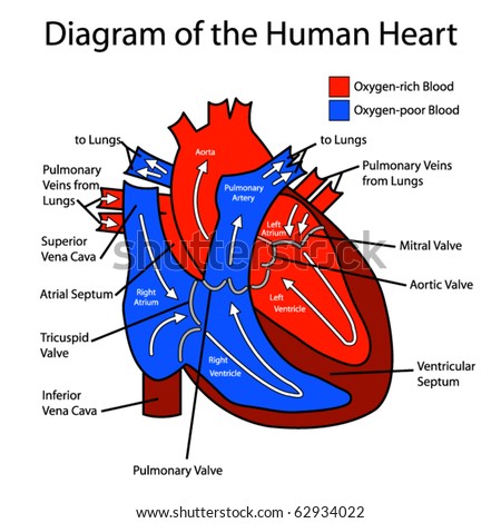
Human Heart
Atria- heart chamber that receives blood
- is thin walled because it only pumps blood to ventricle
Ventricle- heart chamber that pumps blood away from heart to body and back
- therefore thick walled
Valves- prevent back flow of blood
4 chambers = O2 rich blood and O2 poor blood separate
Blood pathway!
- right ventricle to lungs (O2 poor) -> pulmonary (semi-lunar valve) ->
- pulmonary arteries ->
- capillaries O2 diffuses into blood and CO2 diffuses out of blood ->
- pulmonary veins ->
- Left Atrium (O2 rich) -> Bicuspid valve/ mitral valve ->
- Left ventricle ->Aortic valve (semi-lunar valve) ->
- Aorta (largest blood vessel in body) ->
- Capillaries O2 diffuses into tissues, CO2 diffuses out of tissues ->
- Superior and inferior vena cava ->
- R. atrium ->Tricuspid valve -> back to top

Cardiac Cycle
-cardiac cycle- rhythmic contraction (Diastole) and relaxation (Systole) of heart
- a healthy heart rate for an adult is 60-80 beats per minute
- blood moving from the atria to ventricle is the 'ba-dum' sound of the heart
- heart murmur = heart defect
- EKG (electrocardiogram)- uses electrodes to record activity of the heart and can detect the electrical impulses produced by a pacemaker
Controlling the heart rate
- Pacemaker- sets the tempo of the heart beat
- Av (atrioventricular node) delays contracting by 0.1 seconds to ensure atria is emptied completely
- increasing heart rate
- epinepherine (aderenaline)- hormone released during stress, when we were approached by an animal or threat, adrenaline was released to either aid you in running away farther from threat or aid you in fighting off the threat
- caffeine
- exercise
note: higher heart rate = more oxygen to muscles
Notes:
- remember, when looking down at a diagram, you are looking at a patient therefore left and right are switched ex: heart is on the right side of a diagram
- know the pathway of blood inside and out, quiz coming soon (Ms. Andrews said something about a tuesday..?)
- oxygenated blood is red, O2 deficient is blue
- looking back at the unit "The Diversity of Life" might be helpful
Homework: notes, read chapter 23 pg 503-514, try to finish UP 13 (hope the picture below helps if you're stuck)
Next Scribe: Yvette :)
Wednesday, May 9, 2012
May 5
Today we wrapped up the "what happens to the food you eat" unit before the TEST TOMORROW.
First, we took each others quizzes and graded them based on content, appearance, and correctness. It was good review for the test and a reminder of what to study, so if you weren't there, be sure to look through notes and practice questions extra carefully!
After that, we got back our digestion worksheets. If you have any questions regarding the answers, check the table below. (By the way, the intestinal glands make the small intestine enzymes, the large intestine absorbs vitamin K).
and here our our bffs the reagents
We also got our frog labs back.
Then, we checked the answers for our food lab. The results are as follows. Use your creativity for the analysis questions.
The last thing we did was check the second half of the practice quiz we took a couple days ago.
First, we took each others quizzes and graded them based on content, appearance, and correctness. It was good review for the test and a reminder of what to study, so if you weren't there, be sure to look through notes and practice questions extra carefully!
After that, we got back our digestion worksheets. If you have any questions regarding the answers, check the table below. (By the way, the intestinal glands make the small intestine enzymes, the large intestine absorbs vitamin K).
Enzyme/ Substance
|
Organ(s)
|
Works on…
|
Result
|
Salivary Amylase
|
1. Salivary glands
2. Oral cavity/ mouth
|
Starch
|
Maltose
|
Pepsin
|
Stomach
|
Proteins
|
Amino Acids
|
Trypsin
|
Pancreas
|
Proteins
|
Amino Acids
|
Peptidase
|
Small Intestine
|
Proteins
|
Amino Acids
|
Maltase
|
Small Intestine
|
Maltose
|
Glucose
|
Sucrase
|
Small Intestine
|
Sucrose
|
1. Glucose
2. Fructose
|
Rennin
|
Stomach
|
Milk proteins
|
Coagulates
|
Lipase
|
Pancreas
|
Fat
|
1. Glycerol
2. Amino Acids
|
Pancreatic Amylase
|
Pancreas
|
Starch
|
Simple sugars
|
Lactase
|
Small Intestine
|
Lactose
|
1. Glucose
2. Galactose
|
Bile
|
1. Liver
2. Gall Bladder
|
Fat
|
Emulsifies
|
HCl
|
Stomach
|
---------------
|
Function:
1. Activates pepsin
2. Kills bacteria
|
and here our our bffs the reagents
We also got our frog labs back.
Then, we checked the answers for our food lab. The results are as follows. Use your creativity for the analysis questions.
Starch
|
Simple Sugars
|
Protein
|
Fat
|
|
Carrot Juice
|
+
|
+
|
+
|
-
|
Bread
|
+
|
-
|
+
|
+
|
Potato
|
+
|
-
|
+
|
-
|
Apple Juice
|
-
|
+
|
-
|
-
|
Onion
|
-
|
+
|
-
|
-
|
Walnut
|
-
|
-
|
+
|
+
|
Shredded Cheese
|
-
|
+
|
+
|
+
|
Cream Cheese
|
-
|
+
|
+
|
+
|
Honey
|
-
|
+
|
-
|
-
|
Gelatin
|
-
|
+
|
+
|
-
|
The last thing we did was check the second half of the practice quiz we took a couple days ago.
1. Positive
2. Negative
3. C
4. D
5. C
6. Reabsorption
7. Small
intestine, Pancreas
8. C
10. Bile,
lipase
11. Vegetarians
need to be sure to get enough protein, 8 essential amino acids, iron
Thats it! HW: STUDY, have your packet ready tomorrow. Good luck!:)----->
Next scriber: Vinise!!!! (neigh)
Tuesday, May 8, 2012
May 8th, 2012
Today in class: For the entire period, we worked on the What Nutrients are in Foods? lab (which is a separate packet from our regular unit packet). Mrs. Andrews also collected our "Design a Quiz" projects.
As far as homework goes: ~ Finish the nutrient lab that we worked on in class today
~ Work on review packets (TEST this thursday)
NEXT SCRIBE: Emily
As you probably all know, the purpose of the Nutrient lab was to test different foods (about 10 of them) for the presence of macromolecules (particularly starch, simple sugars, protein, and fats or lipids). Because of the many different parts to this lab, your group of four (which were predetermined) had to divide up the work, so one person would find the results for each test, all working at the same time. At the end of the period, the individual groups met with each other and exchanged data from each test.
In part 1 of the lab, your job was to set up the control for the various experiments. The iodine test was first, where you observed the color change when with the mixtures starch and water, water and Lugol's iodine, and starch, water, ad Lugol's iodine all together. If the mixture turned DARK PURPLE, that would be an indication that simple or complex starches were present (as it should have happened with the mixture of all three substances in this first experiment--the rest of the tests should have provided results that indicated there was no starch present).
To test for simple sugars, you had four different test tubes, two contained the mixture of glucose and Benedict's solution, and two contained the mixture of water and Benedict's solution. One of each mixture was to be heated, and the other two were just supposed to be mixed together and observed. Every one of the test tubes should have provided you with a negative result EXCEPT the test tube that had glucose and Benedict's solution that was heated--the color of that particular mixture should have been red, testing positive for glucose.
Next, you had to test for proteins. The three test tubes in this experiment had the mixtures egg albumen, egg albumen and Biuret reagent, and water and Biuret reagent. If the solution turned PURPLE, then this was an indicator for the presence of protein (the rest should have had negative results).
Lastly, you had to test for lipids. One square of an unglazed brown paper bag should have been labeled "oil" and the second, "water." You then had to wait until each substance evaporated to see whether or not the paper was translucent (light can go through, but one cannot see a clear image through the translucent substance)--if it was, lipids were present. If not, than there were no lipids in the substance (distilled water tested negative, the oil tested positive).
After all of these "control tests," we had to perform the same experiments to test foods for each of the four different macromolecules (9 listed substances, and 1 from home).
To review, Benedict's test is an indicator for simple sugars, Biuret's test is an indicator for proteins, Lugol's iodine solution shows the presence or absence of starches, and an unglazed paper bag can be used to test for lipid (fats).
Think about this:
Which of the four macromolecules from the lab could be present in this chicken dinner?
This is basically what we did in the lab today--just think about what is in the food we eat and how the digestive system breaks it down/absorbs its nutrients! :)
As far as homework goes: ~ Finish the nutrient lab that we worked on in class today
~ Work on review packets (TEST this thursday)
NEXT SCRIBE: Emily
As you probably all know, the purpose of the Nutrient lab was to test different foods (about 10 of them) for the presence of macromolecules (particularly starch, simple sugars, protein, and fats or lipids). Because of the many different parts to this lab, your group of four (which were predetermined) had to divide up the work, so one person would find the results for each test, all working at the same time. At the end of the period, the individual groups met with each other and exchanged data from each test.
In part 1 of the lab, your job was to set up the control for the various experiments. The iodine test was first, where you observed the color change when with the mixtures starch and water, water and Lugol's iodine, and starch, water, ad Lugol's iodine all together. If the mixture turned DARK PURPLE, that would be an indication that simple or complex starches were present (as it should have happened with the mixture of all three substances in this first experiment--the rest of the tests should have provided results that indicated there was no starch present).
To test for simple sugars, you had four different test tubes, two contained the mixture of glucose and Benedict's solution, and two contained the mixture of water and Benedict's solution. One of each mixture was to be heated, and the other two were just supposed to be mixed together and observed. Every one of the test tubes should have provided you with a negative result EXCEPT the test tube that had glucose and Benedict's solution that was heated--the color of that particular mixture should have been red, testing positive for glucose.
Next, you had to test for proteins. The three test tubes in this experiment had the mixtures egg albumen, egg albumen and Biuret reagent, and water and Biuret reagent. If the solution turned PURPLE, then this was an indicator for the presence of protein (the rest should have had negative results).
Lastly, you had to test for lipids. One square of an unglazed brown paper bag should have been labeled "oil" and the second, "water." You then had to wait until each substance evaporated to see whether or not the paper was translucent (light can go through, but one cannot see a clear image through the translucent substance)--if it was, lipids were present. If not, than there were no lipids in the substance (distilled water tested negative, the oil tested positive).
After all of these "control tests," we had to perform the same experiments to test foods for each of the four different macromolecules (9 listed substances, and 1 from home).
To review, Benedict's test is an indicator for simple sugars, Biuret's test is an indicator for proteins, Lugol's iodine solution shows the presence or absence of starches, and an unglazed paper bag can be used to test for lipid (fats).
Think about this:
Which of the four macromolecules from the lab could be present in this chicken dinner?
![]() |
This is basically what we did in the lab today--just think about what is in the food we eat and how the digestive system breaks it down/absorbs its nutrients! :)
Monday, May 7, 2012
May 7th, 2012
Today in class we took some practice quizzes and discussed the calorimetry lab.
Homework
- finish calorimetry lab, due tomorrow
- design a quiz, due tomorrow
- study for Tissues, Excretion and Digestion test, Thursday
- make sure you've read the chapters (21 and 22)
Digestion Notes
There are 4 stages of digestion.
- Ingestion: eating food
- Digestion: breaking down the food you eat into small molecules that your body can use
- Absorption: the cells in the lining of the digestive tract take in the molecules and transport them throughout the body using the bloodstream
- Elimination: getting rid of undigested wastes
There are 2 types of digestion.- Mechanical Digestion: physical processes like chewing and stomach churning
- Chemical Digestion: breaking down food with enzymes
- Hydrolysis: breaking down of polymers into monomers using the atoms from water
- Hydrolases: digestive enzymes that catalyze hydrolysis
- Enzymes: specific molecules that start chemical reactions
Mouth (oral cavity)
- chewing, lubricates food, swallowing into the pharynx (first part of the throat, before the split between the trachea and esophagus
- attached to the mouth are 3 pairs of salivary glands: release saliva (1 liter a day) and salivary amylase
- salivary amylase: breaks starches into sugars (maltose)
- muscular tube, uses muscle contractions called peristalsis to push food into the stomach
- no enzymes
- stores food for 2 to 6 hours, can hold 2 liters of food and water
- muscular, churns food and gastric acid into a mixture called acid chyme
- contains gastric juice: pH of .8 to 2 (VERY acidic), hydrochloric acid (HCl) that kills bacteria and activates pepsin, pepsin: breaks proteins into amino acids, rennin: breaks down milk proteins
- stomach lining coated in mucus to protect itself from gastric juice, replaced every 2 to 3 days
- top of the stomach is closed off by the cardiac sphincter (esophageal sphincter in the picture) and the end of the stomach is closed off by the pyloric sphincter
- sphincter: muscular ring that contricts a part of the body until materials need to exit or enter
- produces bile: emulsifies fats, breaking big fat droplets into little fat droplets so they are accessible to enzymes
- bile is stored, concentrated and released by the neighboring gall bladder
- bile is secreted into the duodenum
- pancreatic juice: contains trypsin, lipase and amylase, neutralizes stomach acids in the duodenum
- trypsin: breaks down proteins into amino acids
- lipase: breaks down fats (lipids) into glycerol and fructose
- pancreatic amylase: breaks down starches into sugars (maltose)
- pancreatice juice is secreted into the duodenum
- 2.5 centimeters wide, 6 meters long
- duodenum: first part of the small intestine, mixes pancreatic juice, bile and intestinal juice, 90% of digestion
- peptidase: breaks down proteins into amino acids
- maltase: breaks down maltose (disaccharide) into 2 glucose molecules (monosaccharide)
- sucrase: breaks down sucrose (disaccharide) into glucose (monosaccharide) and fructose (monosaccharide)
- lactase: breaks down lactose (disaccharide) into glucose (monosaccharide) and galacatose (monosaccharide)
- small intestine completes digestion and does most of the aborption
- villi: tiny projections on the lining of the intestine, increase surface area for absorption, absorbs nutrients and passes them into the sorrounding capillaries
- 5 centimeters wide
- colon: majority of large intestine, absorbs water and excess salts
- appendix: small projection where the small and large intestines meet, no purpose in humans, possible function: to store extra "good" bacteria that the digestive system needs?
- rectum: end of the large intestine, stores wastes (feces)
- anus: final sphincter fo the digestive system
Pictures
http://science.howstuffworks.com/environmental/life/cellular-microscopic/cell2.htm
http://en.wikipedia.org/wiki/Mouth
http://www.umm.edu/patiented/articles/stomach_000329.htm
http://www.emedicinehealth.com/pancreatitis/article_em.htm
http://missinglink.ucsf.edu/lm/IDS_106_LowerGI/Lower%20GI/mainpages/smallintestine.htm
Subscribe to:
Posts (Atom)